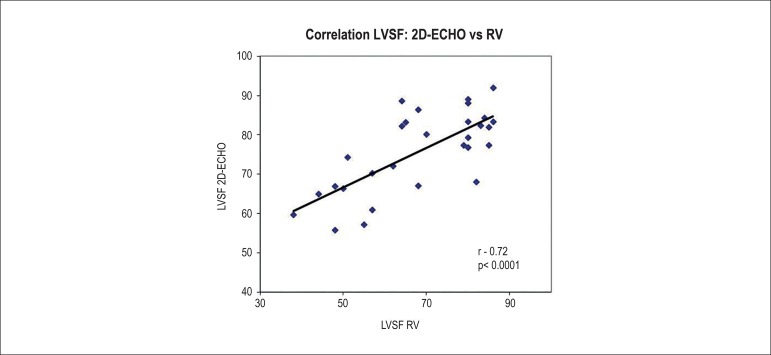
Figure 4.
Graph correlating the left ventricular systolic function (LVSF) assessed by two-dimensional echocardiography (2D-ECHO) and radionuclide ventriculography (RV) in animals receiving different doses of doxorubicin (r = 0.72, p < 0.0001). (*Statistical test performed: linear regression analysis and Pearson's correlation coefficient).

