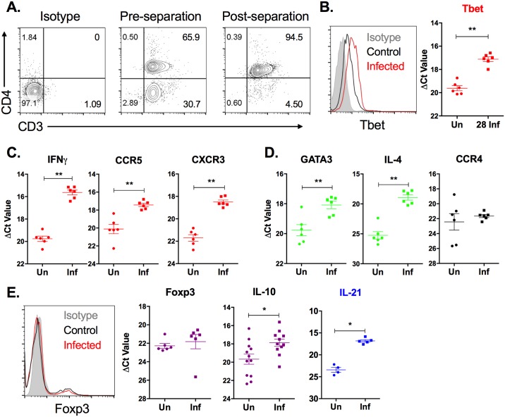
Fig 3. Splenic CD4+ T cell expression of markers of T cell subpopulations over the course of chronic L. donovani infection.
CD4+ T cells were isolated from spleen tissue from uninfected (Un) or 28-day infected (Inf) hamsters by positive selection. (A) The post-separation purity of CD3+CD4+ T cells was >90% in multiple independent experiments. (B-E) RNA was isolated from the purified splenic CD4+ T cell population and mRNA expression of markers of Th1 (B, C), Th2 (D), and Treg cells (E) was determined by real time RT-PCR. Results are expressed as a relative fold-increase between experimental samples and uninfected BHK cells. Shown is the mean and SEM of a single experiment representative of 2 independent experiments from 6 hamsters per group. Expression of (B) T-bet and (E) Foxp3 was verified in CD4+ splenocytes by flow cytometry. Data is representative of at least 2 independent experiments. *p<0.05, **p<0.01. Th1-associated genes indicated by the color red, Th2-associated genes indicated by the color green, Treg-associated genes indicated by the color purple, Th2/Treg-associated genes indicated by the color black, Th1/Th2-associated genes indicated by the color blue.

