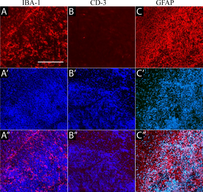
Fig 5. Immunohistochemical characterization of the tumor microenvironment.
(A) Staining against IBA-1 detected microglial activation throughout the tumor. (B) Immunohistochemistry against the T cell marker, CD3, detected very low infiltration, with only single cells observed inside or at the periphery of the tumor. (C) GFAP immunoreactivity was detected in the tumor with all cells expressing high levels of this protein, as well as in the area around the tumor, consistent with activation of surrounding endogenous astrocytes.

