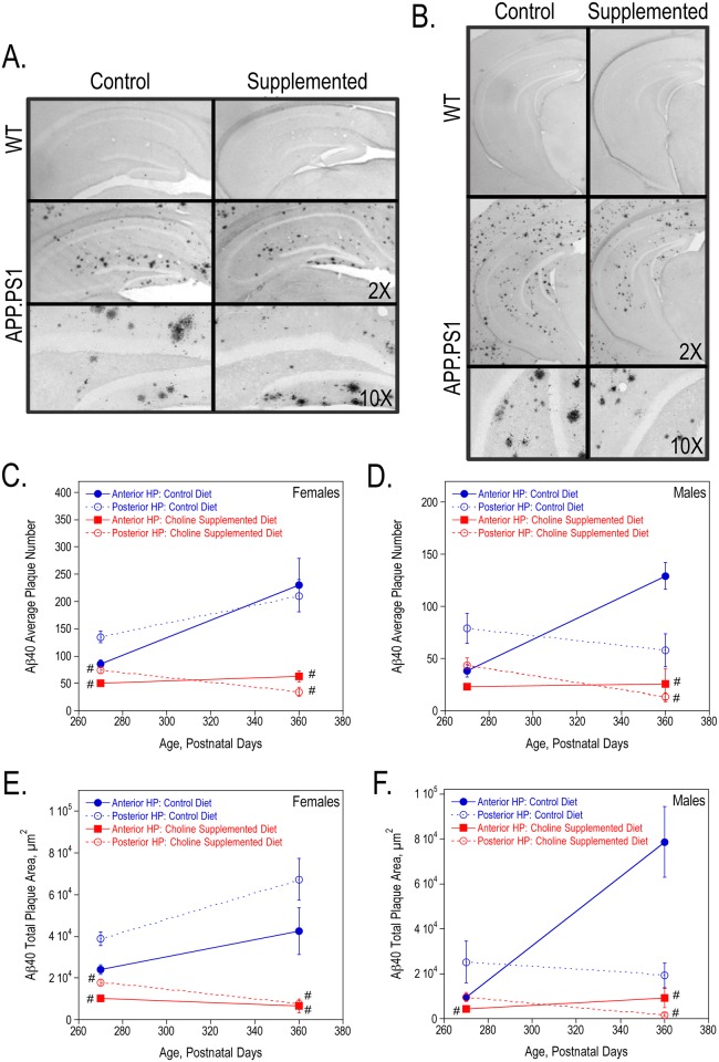
Fig 3. Aβ40 plaques in the hippocampus of 9-, and 12-month old WT and APP.PS1 mice.
Anterior (A) and posterior (B) hippocampal sections from representative 9-month females stained with anti-Aβ40. The average number of Aβ40 plaques per animal (C, D) and the total Aβ40 plaque area (E, F) were quantified using ImageJ64 software in both females and males. As determined by 2-way ANOVA for hippocampal region and diet and Tukey test per age, # represents p<0.05 compared to control diet APP.PS1 mice at the same age. There was a significant overall effect of choline supplementation on the average number and total plaque area in both females (P270: average number p<0.001 and total plaque area p<0.0005; P360: p<0.0001 and p<0.0001) and males (P270: p<0.05 and p<0.05; P360: p<0.0005 and p<0.01).

