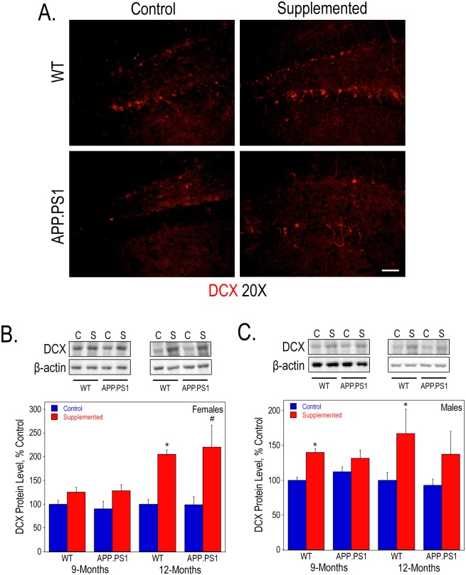
Fig 6. Neurogenesis in the hippocampus WT and APP.PS1 mice.
DCX immunofluorescence staining of anterior hippocampal sections (A) from 9-month old female mice visualized using confocal microscopy. Bar represents 50 μm. Hippocampal lysates of 9- and 12-month old females (B) and males (C) were used to measure DCX protein levels by Western blot analysis. As determined by 2-way ANOVA for genotype and diet and Tukey test per age: * represents p<0.05 compared to control diet WT mice at the same age; and #, p<0.05 compared to control diet APP.PS1 mice at the same age. There was a significant overall effect of perinatal choline supplementation on DCX protein levels in both 9- and 12-month females (p<0.05 and p<0.0005, respectively) and males (p<0.001 and p<0.01, respectively).

