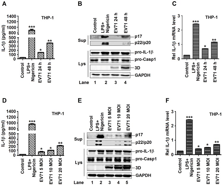
Fig 1. The production and secretion of IL-1β are induced by EV71 in macrophages.
(A, B and C) TPA-differentiated THP-1 cells were infected by the EV71 virus by a time dependent (MOI = 20) for 24 h and 48 h. Supernatants were analyzed by ELISA for IL-1β secretion (A). Immunoblot analysis of the mature (p17) form of IL-1β and cleaved caspase-1 were analyzed in the supernatants (Sup). Cell lysates were normalized for protein content and analyzed by immunoblotting using antibodies specific for pro-IL-1β, pro-caspase-1, EV71 3D, and GAPDH (B). The mRNA levels for the gene pro-IL-1β were quantified by real-time PCR (C). (D, E and F) TPA-differentiated THP-1 cells were infected by the EV71 virus by a dose dependent (MOI = 5, 10, 20) for 24 h. Using the LPS (1 μg/ml) for 6 h and 2 μM Nigericin for 30 min as a positive control. Supernatants were analyzed by ELISA for IL-1β secretion (D). Immunoblot analysis of the mature (p17) form of IL-1β and cleaved caspase-1 in the supernatants (Sup). Cell lysates were normalized for protein content and analyzed by immunoblotting using antibodies specific for pro-IL-1β, pro-Caspase-1, and EV71 3D protein and GAPDH (Lys) (E). The mRNA levels for the gene pro-IL-1β were quantified by real-time PCR (F). Data shown are means ± SEM, *p<0.05, **p<0.01, ***p<0.0001.

