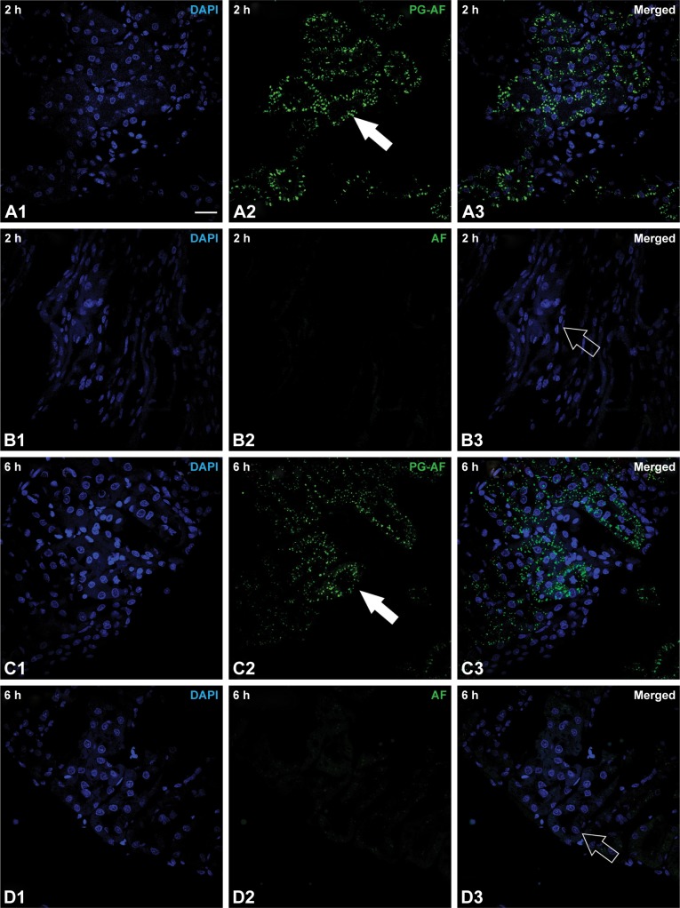
Figure 4.
Histology of normal rat renal tissues at (A) 2 h post 41 kDa PG-AF, (B) 2 h post free AF, (C) 6 h post PG-AF, and (D) 6 h post free AF treatment. 1 Blue fluorescence (refers to nucleus stained with DAPI); 2 green fluorescence (represents AF compound); 3 depicts merged images of 1 and 2. White scale bar at right bottom of A1 =20 µm. PG-AF (filled arrow) was found to accumulate in the epithelial cells of the proximal tubules at 2 h and 6 h posttreatment (A2 and C2). No AF fluorescence (hollow arrow) was detected after 2 and 6 h posttreatment (B3 and D3).
Abbreviations: PG-AF, poly-l-glutamic acid-5-(aminoacetamido) fluorescein (fluoresceinyl glycine amide); DAPI, 4′,6-diamidino-2-phenylindole⋅2HCl; AF, 5-(aminoacetamido) fluorescein.

