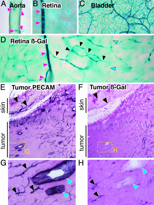
Fig. 4.
Dll4 reporter gene expression reveals prominent expression in smaller arteries and capillaries but not venous vessels in normal adult tissues, with apparent induction during tumor angiogenesis. (A) Whole-mount views of Dll4 reporter expression in aorta and its intercostal artery branches (red arrowheads) of a viable Dll4Lz/+ adult animal. (B) Side-by-side views (from retinal whole mounts) of a small artery (red arrowheads) next to a small vein (blue arrowheads). (C) Prominent Dll4 reporter gene expression in most tissues (urinary bladder wall is shown) seems to extend from small arteries into their downstream microvascular networks. (D) Dll4 expression in an individual vascular circuit (visualized within wholemounts of the adult retina): small arteries, red arrowheads; capillaries, black arrowheads; and post capillary venules, blue arrowheads. (E) Lower-power views of s.c. tumor sections stained with anti-PECAM-1, demonstrating robust staining in all large and small vessels, both in tumor and overlying skin (arrowheads). (F) In contrast, Dll4 reporter analysis reveals stronger expression in tumor vessels as compared with the vessels in the adjacent skin. (G and H) High-power views of boxed areas in E and F highlighting small tumor vessels (black arrowheads) versus large tumor veins (blue arrowheads).

