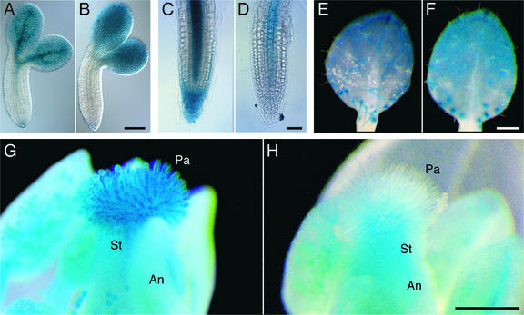
Figure 1.
Histochemical Localization of ADL1A-GUS and ADL1E-GUS Reporter Gene Expression.
(A) and (B) Torpedo-stage embryos.
(C) and (D) Three-day-old seedling root tips.
(E) and (F) Primary leaves of 7-day-old seedlings showing three-branched trichomes.
(G) and (H) Mature flowers (stage 14).
Processing of ADL1A-GUS ([A], [C], [E], and [G]) and ADL1E-GUS ([B], [D], [F], and [H]) tissue samples for the analysis of GUS activity at each developmental stage was performed in a pair-wise fashion. An, anther; Pa, stigmatic papillae cells; St, stigma. Bars = 100 μm in (B) and (D) and 250 μm in (F) and (H).

