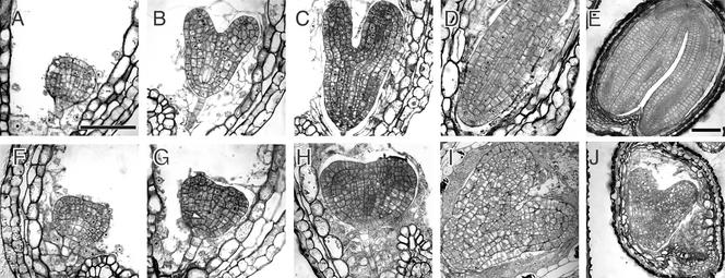
Figure 8.
Development of Homozygous adl1A; E Mutant Embryos.
Bright-field micrographs of histological sections of morphologically wild-type ([A] to [E]) and abnormal homozygous adl1A; E ([F] to [J]) seeds in the siliques of self-fertilized ADL1A/adl1A; adl1E/adl1E plants at various times during development. Embryos in (A) and (F), (B) and (G), (C) and (H), and (D) and (I) are sibling pairs from the same silique. Developmental stages relative to the wild type (Goldberg et al., 1994) are as follows: (A) and (F), globular; (B) and (G), heart; (C) and (H), torpedo; (D) and (I), walking stick; (E) and (J), mature embryo. Mutant embryos were indistinguishable from wild-type embryos before the globular stage. The arrow in (G) indicates mutant cells that failed to expand anisotropically. Bar in (A) = 50 μm for (A) to (D) and (F) to (I); bar in (E) = 100 μm for (E) and (J).

