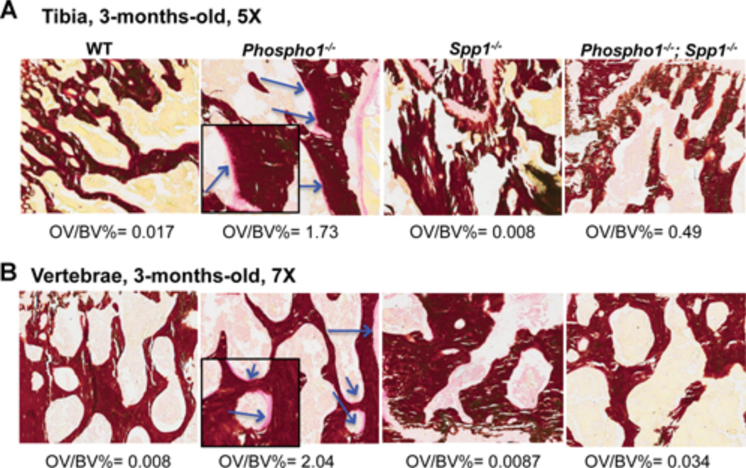
Figure 3.
Histomorphometric analyses of tibias (A) and spines (B) of WT, Phospho1−/−, Spp1−/−, and [Phospho1−/−; Spp1−/−] mice at 3 months of age. Von Kossa/van Gieson staining of the tibial section at the knee joint reveals trabecular bone surrounded by widespread, extended osteoid in 3-month-old Phospho1−/− mice (arrows/inserts show higher magnification of the areas where the osteoid is present), which is less apparent in [Phospho1−/−; Spp1−/−] mice.

