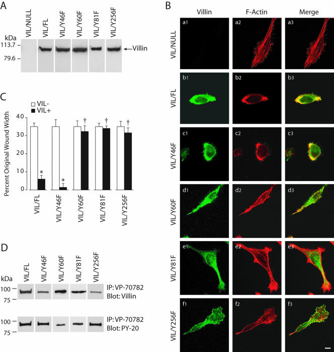
Figure 4.
Tyrosine phosphorylation of villin is required for villin-induced increase in cell migration. (A) HeLa cells were stably transfected with wild-type (VIL/FL) and phosphorylation site mutants of villin, namely, Y46F, Y60F, Y81F, and Y256F. This figure shows representative clones of each villin construct transfected in HeLa cells. Data are representative of six experiments with similar results. (B) Villin expression results in reorganization of the actin cytoskeleton. HeLa cells transfected with wild-type (VIL/FL), and mutant villin proteins were analyzed by confocal microscopy. Double staining of villin (a1–f1) and F-actin (a2–f2) was performed using villin monoclonal antibodies (1:100) and FITC-conjugated anti-mouse IgG (1:200) and Alexa-Phalloidin 568 (1 μg/ml), respectively. Composite images of villin (green) and F-actin staining (red) are shown. Merged images (a3–f3) show colocalization of villin and F-actin. Wild-type villin and VIL/Y46F colocalize with F-actin at the cell periphery. In contrast, phosphorylation mutants of villin VIL/Y60F, VIL/Y81F, and VIL/Y256F show intracellular distribution of villin and F-actin with minimal colocalization of villin and F-actin at the cell surface. Bars, 3 μm. (C) HeLa cells expressing equal amounts of wild-type and phosphorylation site mutants (Y to F) of villin were used in wound-healing experiments. Wounds ∼800 μm in width were made using a pipette tip, and migration of cells into the wound was followed between 0 and 10 h postwounding. Wound repair is expressed as a percentage of the original wound width after 10 h. The error bars are the measured SEM, and the asterisk (*) and cross (†) denote statistically significant values [p < 0.05, n = 24, compared with VIL (-) cells and p < 0.05, n = 24, compared with untreated cells], respectively. VIL (-) refers to each clone cultured in the presence of doxycycline, whereas VIL (+) refers to the same clone cultured in the absence of doxycycline. (D) Tyrosine phosphorylation of wild-type and mutant villin proteins. Cell extracts from VIL/FL and the mutant villin cell lines VIL/Y46F, VIL/Y60F, VIL/Y81F, and VIL/Y256F were immunoprecipitated with phospho-villin antibody (VP-70782) and Western analysis done with villin mAb or phospho-tyrosine antibody (PY-20). This is not a quantitative Western blot. The Western blot is representative of three other experiments with similar results.

