Fig 6.
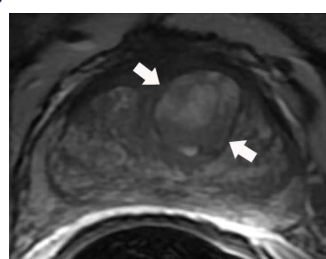
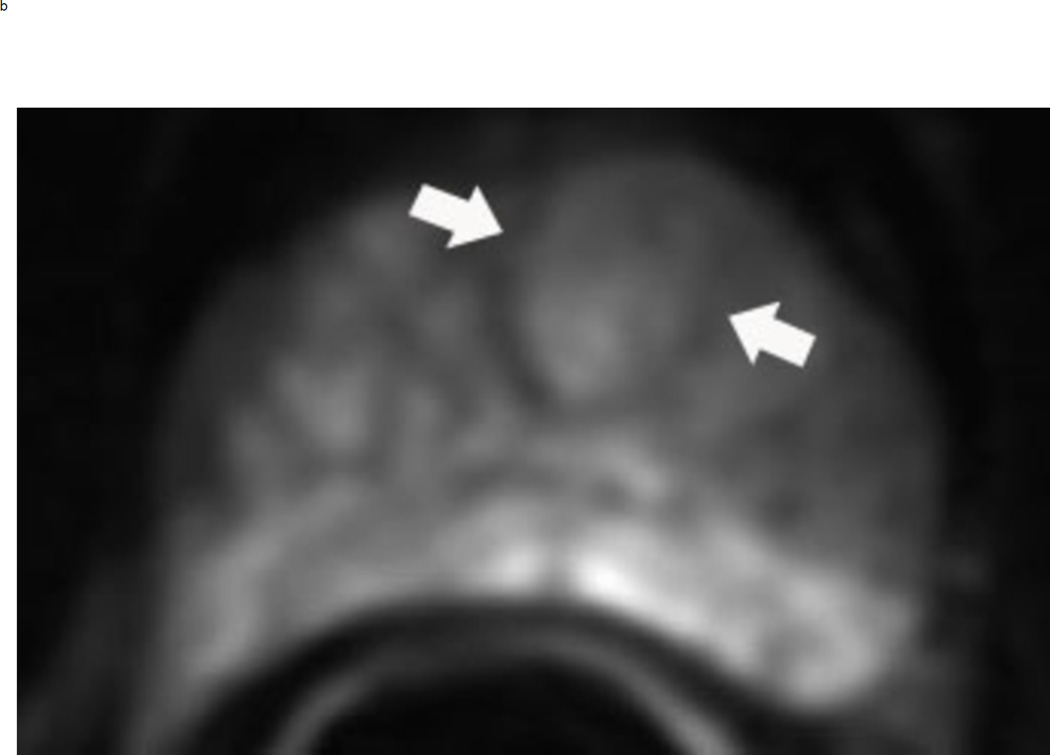
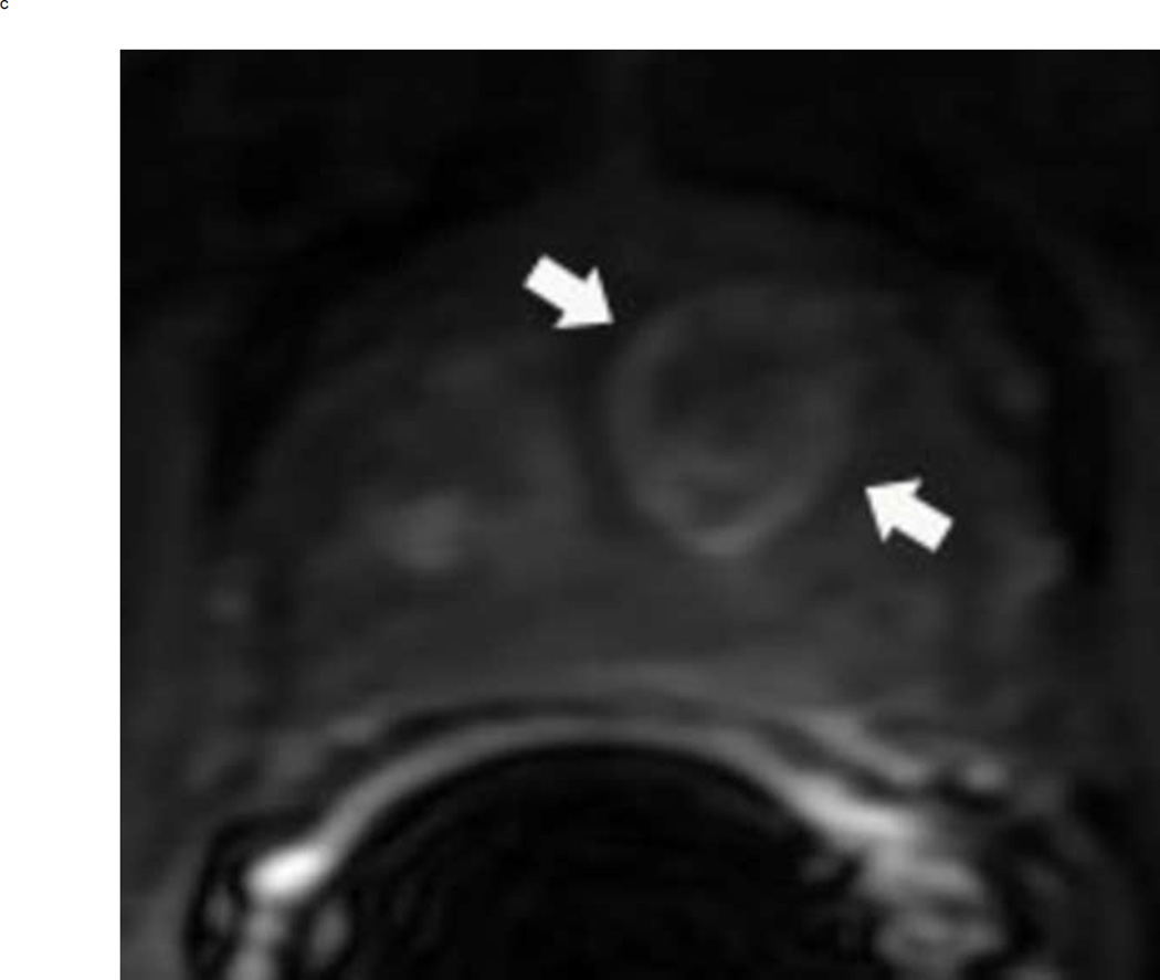
64 year old man with elevated PSA. Axial T2 weighted images show typical appearance of changes of glandular and stromal hyperplasia in the transition zone (arrows) (a). These typical BPH nodules demonstrate mildly increased signal on high b-value diffusion weighted images (arrows) (b) and enhance earlier than the surrounding normal prostatic tissue (arrows) (c). This lesion would be assigned a PI-RADS score of 2. Note: BPH = benign prostatic hyperplasia, PSA = prostate specific antigen.


