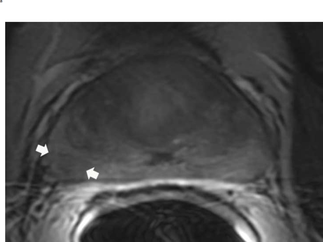
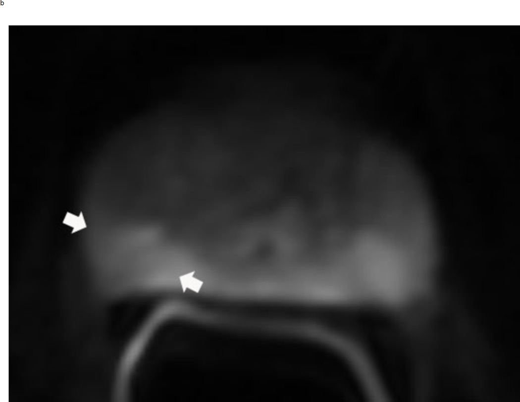
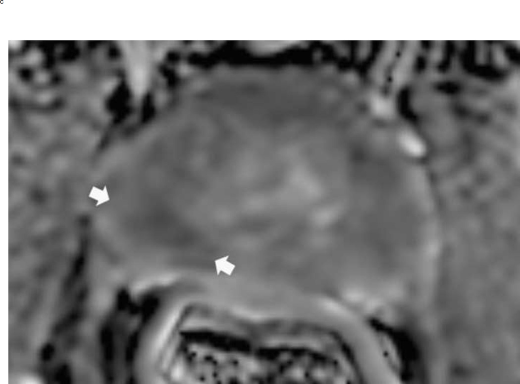
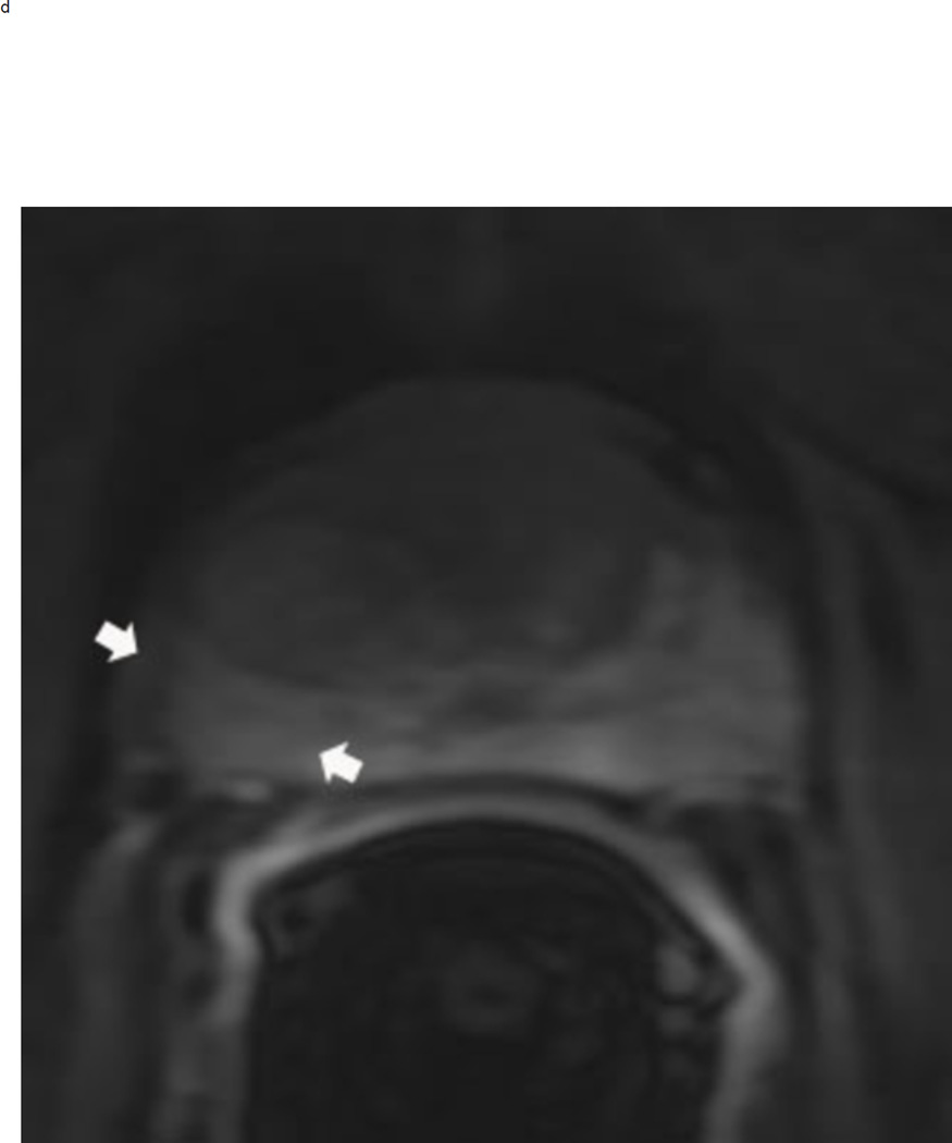
Fig 7.
PI-RADS 3 lesion in the right peripheral zone in a 59 year old man with an elevated PSA. Axial T2 weighted image demonstrates a non-circumscribed rounded moderate signal intensity lesion in the right peripheral PZpl sector (arrows) (a). The lesion is mildly hyperintense on high b-value diffusion weighted image (b) and mildly hypointense on ADC map (arrows) (c). DCE is negative as there is no focal enhancement (arrows) (D). The overall PI-RADS score is 3.


