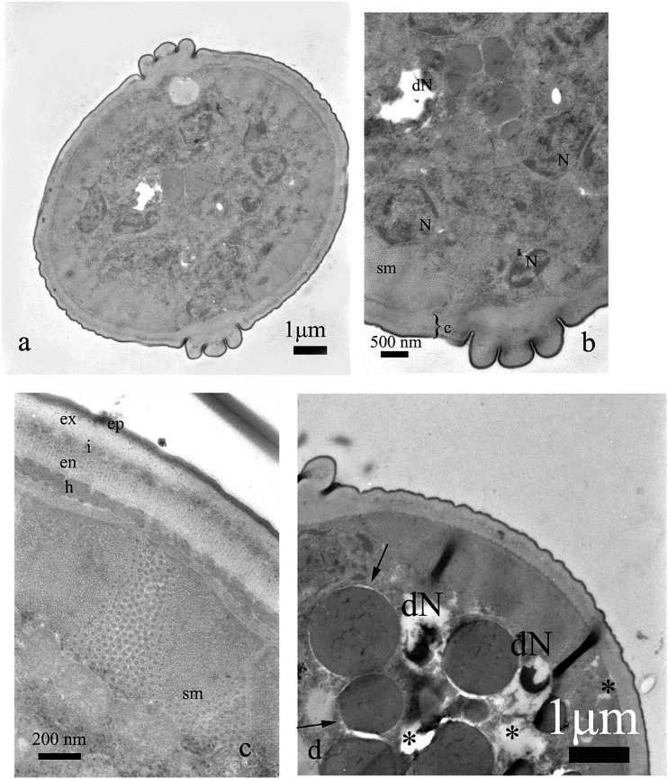
Fig. 6.
Cross section of the nematode treated with undecanone, 20 μg⋅mL−1. Some nuclei were destroyed (dN, b, d), whereas the majority of cells were not altered (a, b). Vacuolization (electron lucent areas) was present within cytoplasm (*, d). Cuticle, its sublayers (ep = epicuticle, ex = exocuticle, i = intermediate zone, en = endocuticle), hypodermis (h) and somatic muscles (sm) were not significantly altered, too (c, d).

