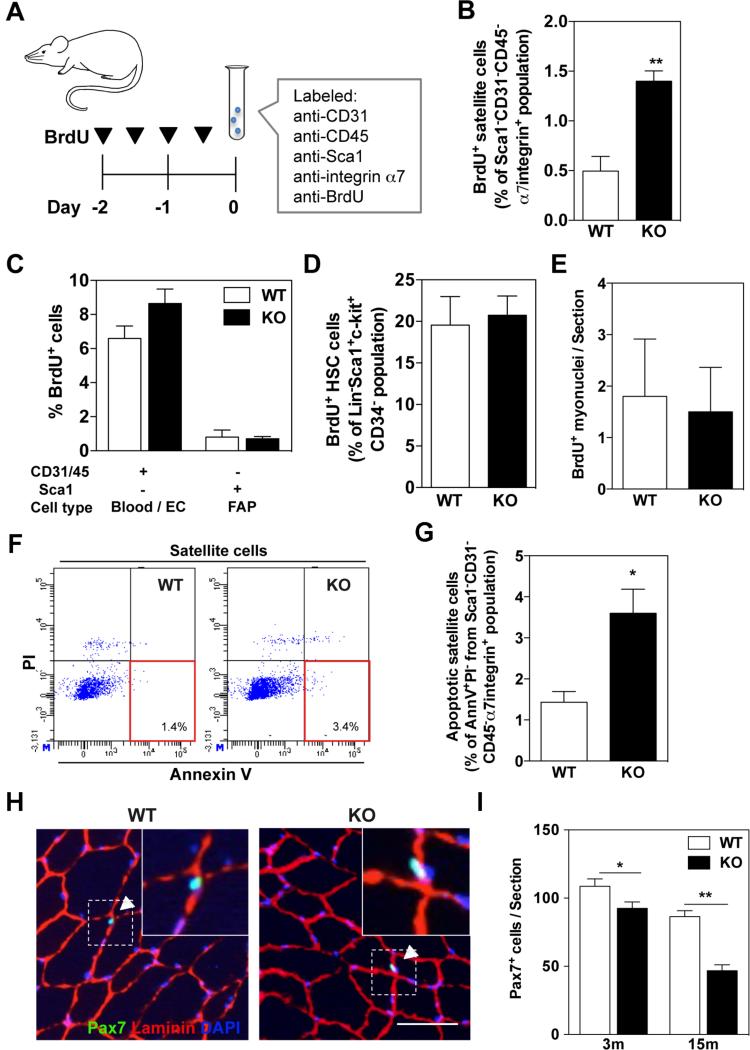
Figure 2. Increased satellite cell proliferation and apoptosis in uninjured muscle s of KPNA1 null mice.
(A) Schematic of BrdU injection and immunolabeling of satellite cells for FACS analysis. (B) The percentage of BrdU+ satellite cells in KPNA1 null (KO) hindlimb muscles was increased approximately 3-fold relative to wild type (WT). (C) The percentage of BrdU+ blood/endothelial cells (EC) and fibro/adipocyte progenitors (FAP) did not significantly differ between WT and KO hindlimb muscles. (D) By flow cytometry, the percentage of BrdU+ hematopoietic stem cells (HSC) did not significantly differ between W T and KO mice. Lin = CD3/Gr-1/CD11b/CD45R/Ter-119 (E) The number of BrdU+ myonuclei in sections of tibialis anterior (TA) muscles did not differ between uninjured WT and KO mice. (F) Representative flow plots depicting apoptotic satellite cells positive for Annexin V and negative for propidium iodide (PI). (G) The percentage of apoptotic satellite cells was increased approximately 2.5-fold in KO hindlimb muscles relative to WT. AnnV = Annexin V. (H) Representative images of TA muscles immunostained for Pax7 (green), laminin (red), and DAPI (blue). Bar = 50 μm. White arrowheads indicate Pax7+ satellite cells co-stained with DAPI and located inside of laminin outlines. (I) Pax7+ satellite cells were significantly decreased in TA muscles of KO mice relative to W T at both 3 and 15 months of age. Data represent the mean ± SEM. n=3 for all experiments. **p<0.01 and *p<0.05.

