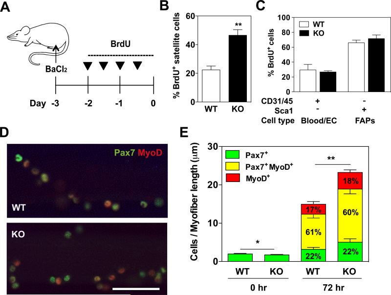
Figure 3. Increased satellite cell proliferation in regenerating muscles of KPNA1 null mice.
(A) Schematic of muscle injury induced by BaCl2 and BrdU injection protocol. (B) The percentage of BrdU+ satellite cells in regenerating KPNA1 null (KO) gastrocnemius muscles was increased about 2-fold relative to wild type (WT). (C) The percentage of BrdU+ blood/endothelial cells (EC) and fibro/adipocyte progenitors (FAPs) did not significantly differ between regenerating gastrocnemius muscles of WT and KO mice. (D) Representative images of WT and KO myofibers immunostained for Pax7 (green) and myoD (red) after 72 hours of culture. Bar = 50 μm. (E) Pax7+, Pax7+MyoD+ and MyoD+ satellite cells were quantified on myofibers at 0 and 72 hours of culture in order to analyze lineage progression. The total number of KO satellite cells was increased by approximately 30% after 72 hours of culture. The percentage of satellite cells at each stage of lineage progression did not significantly differ between WT and KO myofibers. WT myofibers=15; KO myofibers=21. Data represent the mean ± SEM. n=3-5 for all experiments. **p<0.01 and *p<0.05.

