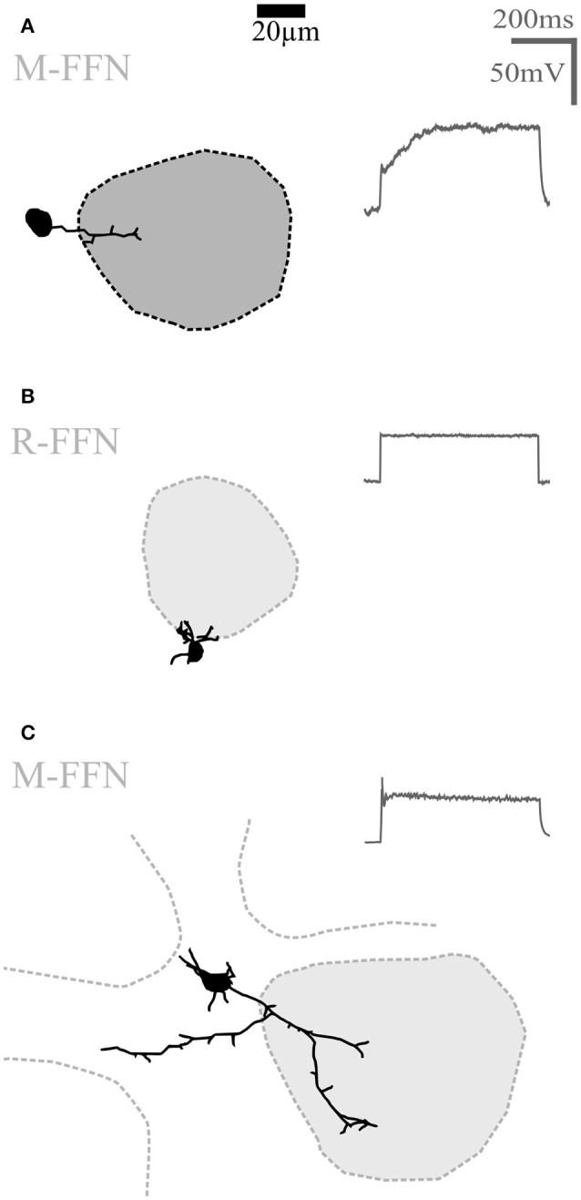
Figure 4.

Undersized cells (UCs). Maximal z-projections of reconstructions of representative UCs. Shading of glomeruli corresponding to Figures 2, 3. None of the reconstructed UCs showed truncations or signs of bad filling. UCs also never showed regular sodium action potential firing (voltage traces shown on the right in panels A–C) upon step depolarizations. (A) Morphologically very simple cell entering a single glomerulus. (B) Even smaller cell that does not enter the adjacent glomerulus. (C) Slightly larger cell compared to (A,B), extending dendrites in the juxtaglomerular space. R-FFN, FFN102+ cells in juvenile rats; M-FFN, FFN102+ cells in adult mice.
