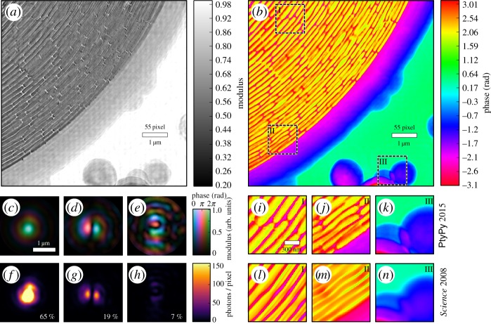
Figure 6.
X-ray ptychography at a small-angle scattering beamline of a synchrotron. (a,b) Recovered modulus and phase of the outer region of a zone plate, serving here as a sample. (c–e) Wavefields of three recovered probe modes that represent 91% of the illumination in the sample plane. The probe modes were orthogonalized with the main mode being the mode to the left (c). (f–h) The same probe modes but their intensity distribution is displayed on a linear scale. Note that the main mode reaches values up to 700 photons in its centre. The relative power of the mode is shown in the bottom right corner. (i–k) Selected regions of the recovered phase (b) displayed alongside the same regions (l–n) from the original phase reconstruction [7] of the sample. An improvement in reconstruction quality is apparent: small bridges for stabilizing the ring structure are visible in (i) but not in (l). Deviations of the outer zones from a circular path can be observed in (j) but can only be guessed from (m).

