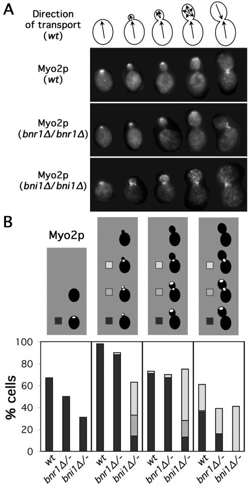
Figure 2.
Myo2p localization is perturbed in bni1Δ cells. (A) Models as in Figure 1A are depicted with selected wild-type (ABY1848), bnr1Δ/bnr1Δ (ABY1801), and bni1Δ/bni1Δ (ABY1867) cells stained to show Myo2p distribution. (B) One hundred cells of each budding category treated as described in A were scored for Myo2p localization. Unbudded cells were categorized as having Myo2p delocalized (blank) or localized to one site on the cell surface (black). Budded cells were scored as having Myo2p localized to the bud cortex (though not necessarily the bud tip) (black), the bud cortex and neck (gray), localized to the bud neck (white), or delocalized (blank).

