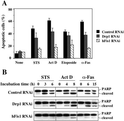
Figure 3.
hFis1 depletion produces greater resistance to apoptosis than depletion of Drp1. (A) HeLa cells depleted of hFis1 or Drp1 by RNAi along with control RNAi cells were treated with STS (1 μM; 6 h), Act D (10 μM; 8 h), etoposide (100 μM; 30 h), or anti-Fas antibody (500 ng/ml, 15 h), and the nuclei were stained with Hoechst 33342 (1 μg/ml; 15 min at RT). Normal or apoptotic nuclei of these cells in several fields were counted under the fluorescent microscope (for UV excitation). At least 200 cells altogether in each treatment were counted and shown as a percentage of cells with apoptotic nuclei among total cells counted. The data are plotted as the mean ± SD of at least three independent experiments. (B) PARP cleavage was analyzed in the total extracts from HeLa cells depleted of hFis1, Drp1, and control, which were treated with STS (1 μM; 0, 3, 6 h), Act D (10 μM; 0, 4, 8 h), or anti-Fas (500 ng/ml; 0, 6, 15 h). The figure is a representative of at least three independent experiments.

