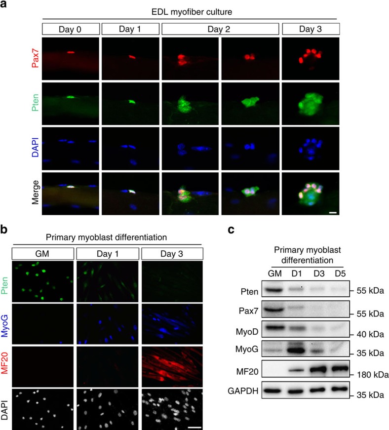
Figure 1. Pten is expressed abundantly in quiescent and activated SCs.
(a) Pten immunofluorescence in Pax7+ SCs attached on freshly isolated EDL myofibers (Day 0) or after cultured for 1–3 days. Scale bar, 10 μm. (b) Co-immunostaining of Pten, MyoG (differentiation marker) and MF20 (myosin heavy chain) in primary myoblasts in growth medium (GM) or differentiated for 1–3 days. Scale bar, 50 μm. (c) Western blot showing relative levels of Pten and myogenic marker proteins at various stages of myogenic differentiation.

