FIG 1.
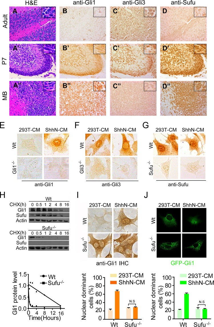
Sufu is essential for the upregulation and nuclear localization of Gli1. Hematoxylin and eosin (H&E) staining (A to A″) and immunohistochemistry staining (B to B″, C to C″, and D to D″) were carried out on adult (6 months old) and developing (P7) cerebellar and medulloblastoma sections. (E to G) IHC staining in wild-type and mutant MEFs. The proportions of cells showing Shh-induced nuclear enrichment are approximately 67.5% (Gli1 [E]), 3% (Gli3 [F]), and 70% (Sufu [G]), respectively. (H) Western analyses showing the turnover rate of Gli1 in wild-type and Sufu−/− MEFs. (I) IHC staining of Gli1 in wild-type and Sufu−/− MEFs. The cells were treated with control (293T cells) or ShhN-conditioned medium (ShhN-CM) for 24 h before they were fixed in 4% PFA. The graphs were calculated based on two independent experiments (n = 100). (J) Immunofluorescence visualization of GFP-Gli1 in transiently transfected MEFs. The cells were treated and the graphs were calculated as for panel I (n = 50).

