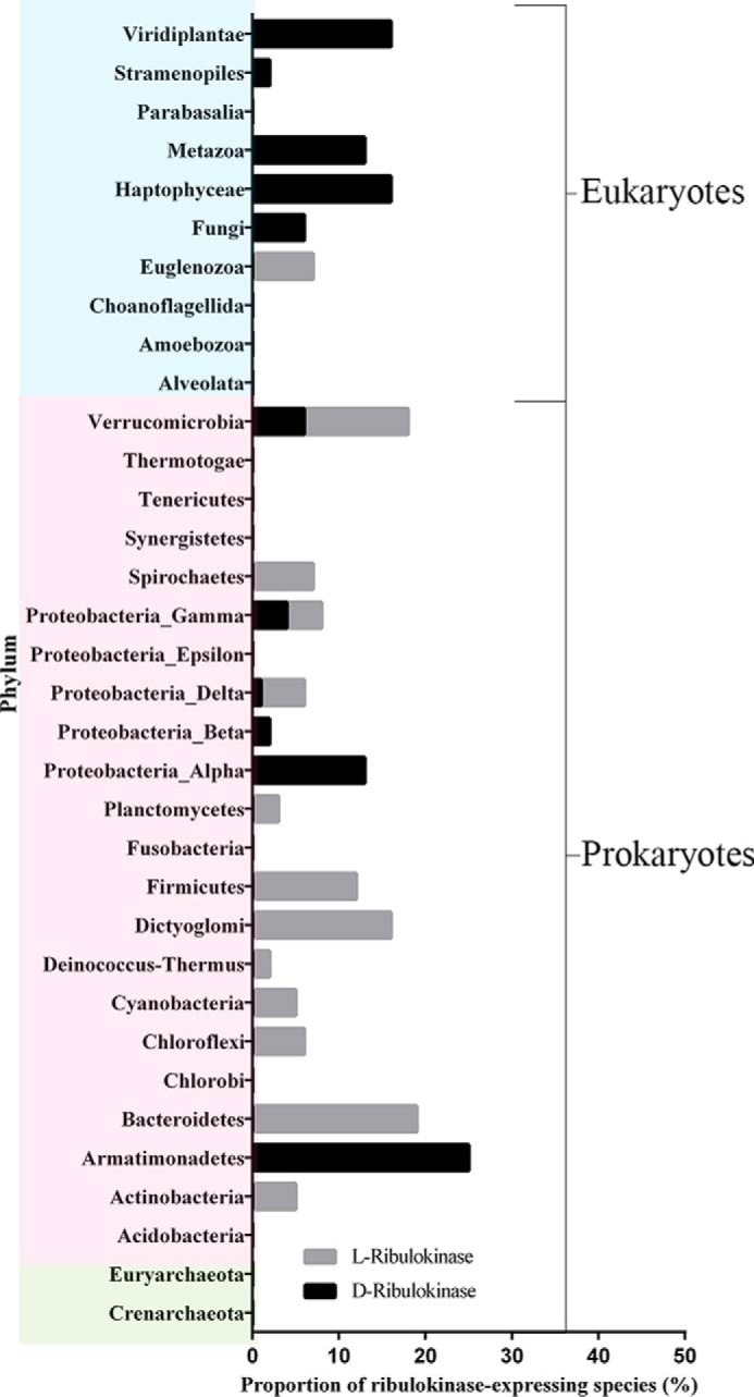
FIGURE 9.

Phyletic spread of d-ribulokinase and l-ribulokinases. The taxonomic distribution of d- and l-ribulokinase proteins was determined using the FGGY_N domain sequences present in the Pfam database at the time of analysis and the d-ribulokinase and l-ribulokinase motifs defined in this study (TCSLV and TGTST, TSST, TGSSP, TGSTP, MMHGY, and TACTM, respectively). Eukaryotic, bacterial, and archaeal phyla are represented on blue, pink, and green backgrounds, respectively.
