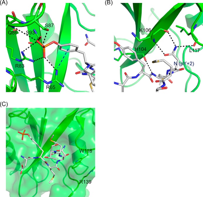
FIGURE 3.

The site-specific recognition modes of Gads SH2. A, the interactions of phosphotyrosine of CD28-derived phosphopeptide with Gads SH2. B, the interactions (salt bridges and hydrogen bonds) of the conserved Asn (pY+2) of CD28-derived phosphopeptide and Gads SH2. C, SH2 domain is shown in ribbon and surface representation, and CD28-derived phosphopeptide is shown in stick representation. The black dashed lines indicate the salt bridges and hydrogen bonds. The blue dashed line indicates cation-π interaction between CD28-derived phosphopeptide and SH2.
