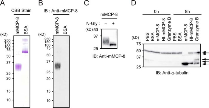
FIGURE 1.
Preparation and characterization of recombinant mMCP-8 protein. A and B, recombinant mMCP-8 protein purified from culture supernatant of mMCP-8-transduced Sf9 cells was resolved by SDS-PAGE, followed by detection with Coomassie Brilliant Blue (CBB) staining (A) or immunoblotting (IB) using an mMCP-8-specific mAb (anti-mMCP-8, B). BSA was used for control experiments. C, purified recombinant mMCP-8 was treated with 100 units/ml N-glycosidase (N-Gly) at room temperature overnight or left untreated, followed by immunoblot analysis using anti-mMCP8 mAb. D, total cell lysates of NIH3T3 cells were incubated at room temperature for 8 h in the presence or absence of the indicated proteins at a concentration of 10 μg/ml. After incubation, the cell lysates were subjected to immunoblot analysis with anti-α-tubulin polyclonal Ab. The bands corresponding to the full-length α-tubulin protein and its degradation products are indicated by arrows. The same set of cell lysates without incubation is shown as 0 h. Data shown are representative of at least three independent experiments.

