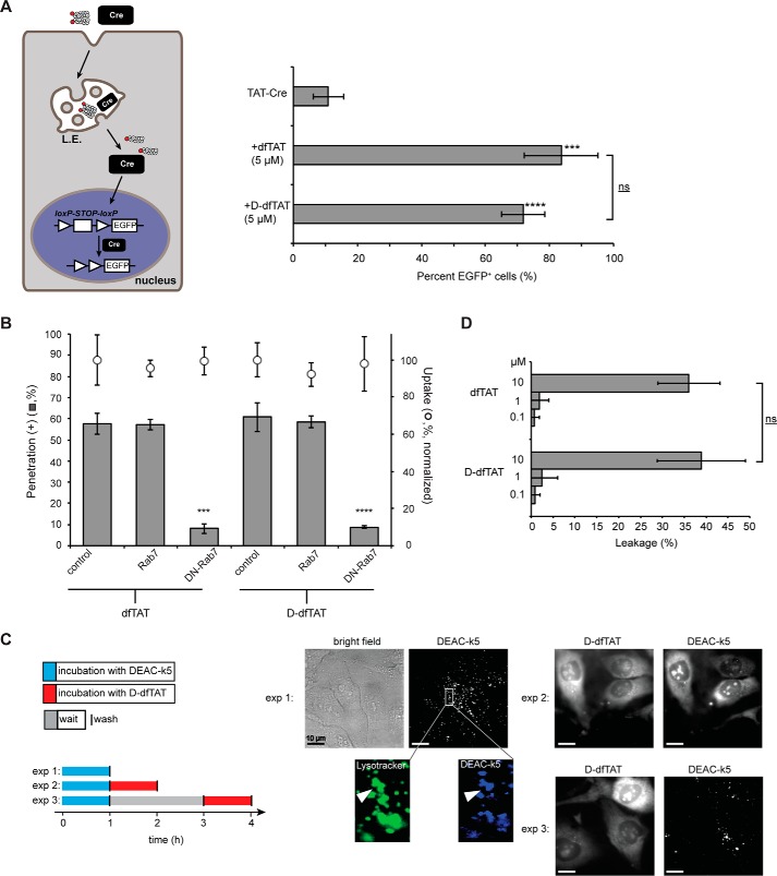
FIGURE 2.
d-dfTAT enters cells by endocytosis followed by endosomal escape in a manner similar to dfTAT. A, dfTAT and d-dfTAT deliver Cre recombinase into live cells. HeLa cells were transfected with a plasmid containing EGFP upstream of a loxP-STOP-loxP sequence. The cells were then co-incubated with dfTAT or d-dfTAT (5 μm) and 4 μm Cre recombinase. Cells positive for EGFP expression were counted 24 h after the protein delivery treatment. ***, p ≤ 0.001; ****, p ≤ 0.0001 compared with control (TAT-Cre). The bracket indicates comparison of p values obtained using t test analysis of dfTAT and d-dfTAT treatments (ns, p > 0.05). B, cytosolic penetration of d-dfTAT is blocked by expression of dominant negative Rab7. HeLa cells were transfected with Rab7 or dominant negative Rab7 (DN-Rab7). dfTAT or d-dfTAT (3 μm) was then incubated with cells for 1 h, and cell penetration was quantified. The total fluorescence of cell lysates was also measured to assess the impact of each treatment on peptide endocytic uptake. The endocytic uptake of cells transfected with Rab7 or DN-Rab7 was normalized to the endocytic uptake of cells treated with dfTAT or d-dfTAT, respectively. ***, p ≤ 0.001; ****, p ≤ 0.0001 compared with control. C, d-dfTAT causes the release of a peptide preloaded into endosomes but not that of a peptide preloaded into lysosomes. In experiment 1 (exp 1), cells were incubated with DEAC-k5 (20 μm) for 1 h and washed. Cells were then incubated with LysoTracker during imaging to establish the accumulation of DEAC-k5 inside endosomes. In experiment 2 (exp 2), cells were incubated with DEAC-k5 (20 μm) for 1 h, washed, and then incubated with d-dfTAT (5 μm) for 1 h. Experiment 3 (exp 3) was performed as experiment 2 with the exception of a 2-h waiting time between incubation with DEAC-k5 and d-dfTAT. Fluorescence images represented are either monochromatic or pseudocolored green for LysoTracker and blue for DEAC-k5. Scale bars, 10 μm. D, d-dfTAT causes leakage of liposomes with a lipid composition mimicking that of late endosomes. Liposomes loaded with calcein (500 μm) were incubated with dfTAT or d-dfTAT (0.1, 1, or 10 μm). The fluorescence signal of free calcein was quantified after peptide treatment. ns, p > 0.05. The data reported in this figure represent the average and corresponding S.D. values (error bars) of biological triplicates.

