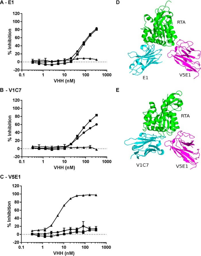
FIGURE 4.
V5E1 recognizes an epitope on RTA that is spatially distinct from E1 and V1C7's epitopes. Competition ELISA in which E1 (A), V1C7 (B), or V5E1 (C), at indicated concentrations, was mixed with biotinylated ricin in solution and then applied to microtiter plates coated with E1 (circles), V1C7 (squares), or V5E1 (triangles). The plates were then probed with avidin-HRP, and the amount that the soluble VHH blocked biotin-ricin capture by the plate bound antibody is indicated on y axis (% inhibition). D and E, structure of RTA-V5E1 complex was superpositioned onto the RTA-E1 or RTA-V1C7 structures to demonstrate the distinct binding profiles. RTA is colored green; V5E1 is magenta, and E1 and V1C7 are in cyan.

