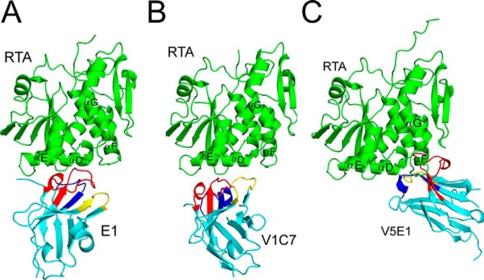
FIGURE 6.
X-ray crystal structures of RTA-VHH complexes. Structures of RTA in complex with VHHs E1 (A), V1C7 (B), and V5E1 (C). RTA (green) is presented in a similar orientation for each panel with α-helices D–G indicated, as necessary. VHHs are shaded in cyan with CDR1–3 colored blue, yellow, and red, respectively.

