FIGURE 4.
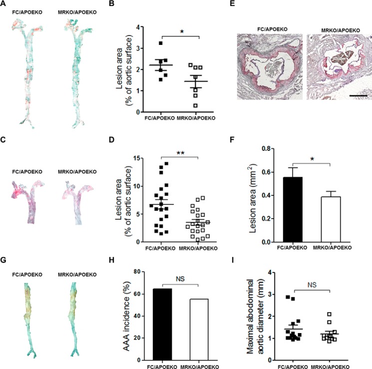
Effects of myeloid MR deficiency on atherosclerosis and abdominal aortic aneurysm in APOEKO mice. A, representative images of en face oil red O staining of aortas from FC/APOEKO mice and MRKO/APOEKO mice infused with saline for 28 days. B, quantification of A as percentage of total aortic surface (n = 6–7). C, representative images of en face oil red O staining of aortas from FC/APOEKO mice and MRKO/APOEKO mice infused with AngII for 28 days. D, quantification of C as percentage of total aortic surface (male, n = 6–8; female, n = 14–12). E, representative images of oil red O staining of the aortic sinus from FC/APOEKO mice and MRKO/APOEKO mice. Scale bar, 500 μm. F, quantification of the atherosclerotic lesion area of the aortic sinus (n = 5). G, representative images of aortic aneurysm in abdominal aortas from FC/APOEKO mice and MRKO/APOEKO mice infused with AngII for 28 days. H, the incidence of abdominal aortic aneurysm in FC/APOEKO and MRKO/APOEKO mice (n = 16–17). I, maximal aortic diameter of abdominal aortas from FC/APOEKO and MRKO/APOEKO mice (n = 11–14). NS, not significant. *, p < 0.05; **, p < 0.01.

