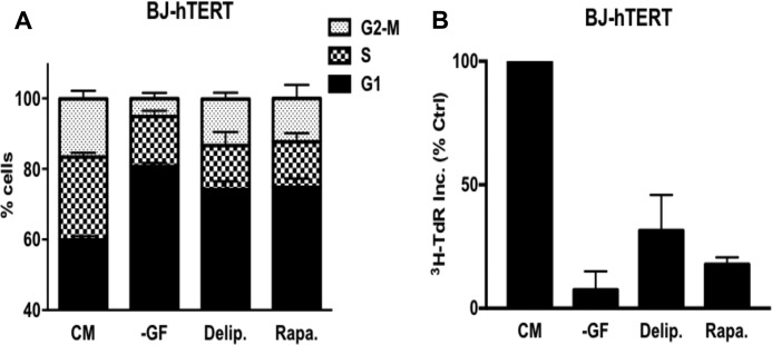
FIGURE 1.
Depriving cells of lipids arrests cells in G1. A, BJ-hTERT cells were plated at 30% confluence in DMEM containing 10% FBS. After 24 h, cells were shifted to complete medium (CM), no growth factors (−GF), medium containing 10% delipidated serum (Delip.), or CM containing rapamycin (Rapa.) for 48 h, after which the cells were harvested and analyzed for cell cycle distribution by measuring DNA content/cell. The CM contained 10% dialyzed FBS instead of 10% FBS. Error bars represent the standard deviation from independent experiments repeated 3 times. B, BJ-hTERT cells were plated and shifted to the conditions explained above. Cells were labeled with [3H]TdR for the final 24 h of treatment, after which the cells were collected and the incorporated label (3H-TdR Inc.) was determined by scintillation counting. Ctrl, control. Values were normalized to the cpm for CM, which was given a value of 100%. Error bars represent the standard deviation for experiments repeated 3 times.

