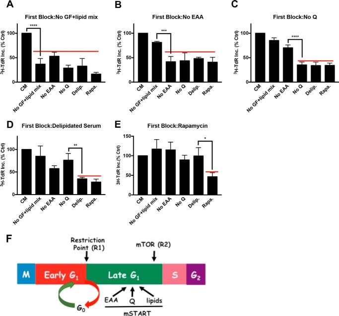
FIGURE 2.
Lipid deprivation arrests cells downstream of the Gln checkpoint and upstream from the mTOR checkpoint. BJ-hTERT cells were plated and shifted to various first blocking conditions for 48 h as in Fig. 1A. The cells were subsequently shifted to CM or different second block conditions containing [3H]TdR for 24 h, after which the cells were collected and the incorporated label (3H-TdR Inc.) was determined. The first blocks were: A, no serum (no growth factors) plus lipid mix (No GF+lipid mix) (see “Experimental Procedures”); B, no EAA; C, no Gln (Q); D, delipidated serum; and E, rapamycin (20 μm). Ctrl, control; No Q, no Gln; Delip., delipidated serum; Rapa., rapamycin. F, schematic summarizing results from the double block mapping experiments for metabolic checkpoints for EAA, Gln, and lipids that are hypothesized to represent a mammalian START (mSTART). Also shown are the relative positions of the two restriction points (R1 and R2) that respond to growth factors that have been described (5, 7). Error bars represent the standard deviation for experiments repeated at least 3 times. One-way analysis of variance was used to generate p values for establishing the significance between the primary block condition and the immediate upstream block condition (*, p < 0.05; **, p < 0.01; ***, p < 0.001; ****, p < 0.0001).

