FIGURE 1.

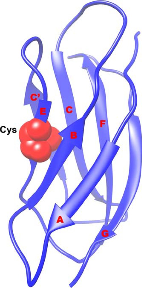
Structure of FNIII 7 showing the seven strands (A, B, C, C′, E, F, and G). The buried Cys in the E strand used in the thiol exchange assay is shown in red. Figure based on the coordinates from 1FNF.pdb (2).

Structure of FNIII 7 showing the seven strands (A, B, C, C′, E, F, and G). The buried Cys in the E strand used in the thiol exchange assay is shown in red. Figure based on the coordinates from 1FNF.pdb (2).