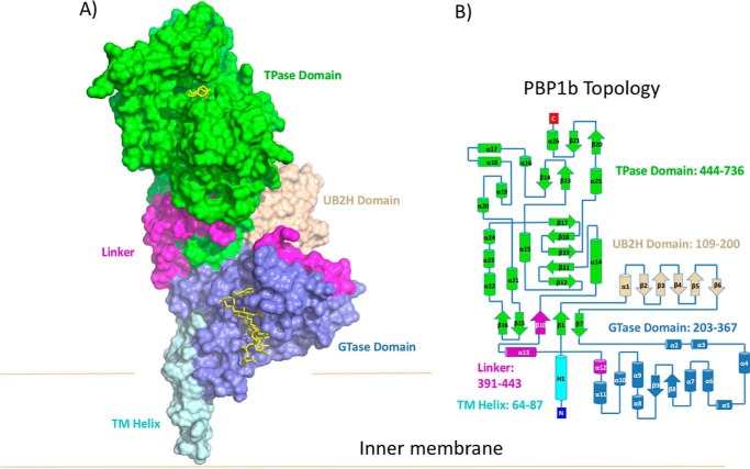
FIGURE 1.
Overall structure of PBP1b-moenomycin-acyl-β-lactam complexes. A, the crystal structure of PBP1b is shown in surface representation. The TM, UB2H, GTase, linker, and TPase domains are colored cyan, beige, blue, pink, and green, respectively. Moenomycin and acyl-ampicillin bound to the GTase and TPase domain are shown in yellow stick representation. B, topology diagram of PBP1b, color-coded as in A.

