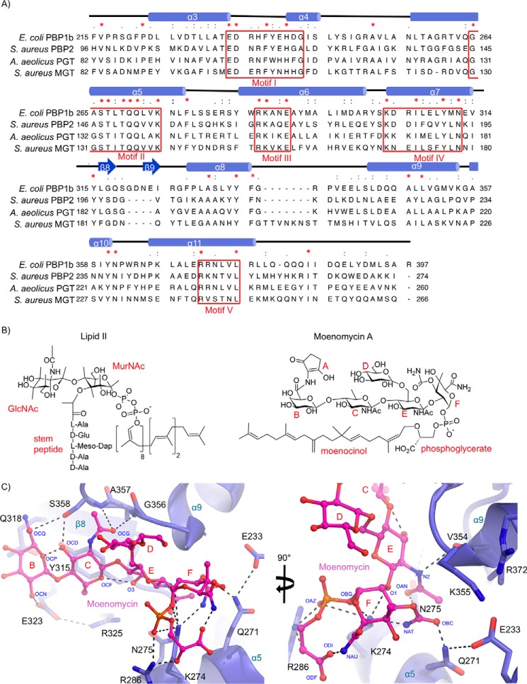
FIGURE 5.
Moenomycin binding to the donor site of the E. coli PBP1b GTase domain. A, multiple sequence alignment of PG GTase domains that have been structurally characterized. The sequences are aligned using tree based progressive alignments and displayed according to their secondary structure elements using ClustalW2 (57) and Chimera (58). B, chemical structures of lipid II and moenomycin. C, donor site close-up of moenomycin-bound to PBP1b. The GTase domain protein backbone is shown in blue cartoon representation with key active site residues displayed as blue sticks with non-carbon atoms colored by type. The bound moenomycin is shown as pink sticks with atoms colored by type. Key hydrogen bonding and electrostatic interactions are shown as black dashes.

