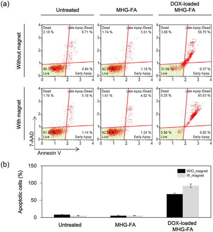
Figure 6. Detection of apoptosis of HeLa cells.
Cells were treated with 100 μg/mL MHG-FA or DOX-loaded MHG-FA for 30 min and further incubated for 48 h. Apoptosis was analyzed by Annexin V/7-AAD double staining and flow cytometry. (a) Representative results from three independent experiments. The lower right quadrant represents early apoptotic cells stained mainly by Annexin V and the upper right quadrant stands for late apoptotic cells stained by both Annexin V and 7-AAD. (b) The percentages of both apoptotic (late and early) and necrotic cells induced by MHG-FA and DOX-loaded MHG FA.

