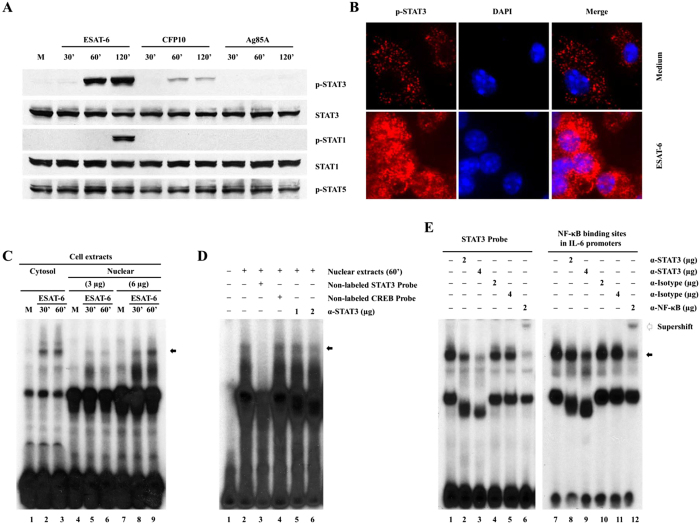
Figure 2. ESAT-6 activates STAT3 in macrophages.
(A) BMDMs were incubated with 1 μg/ml ESAT-6, CFP10 or Ag85A for different periods and the expression of the proteins was determined by Western blotting. One representative result of five independent experiments is shown. (B) BMDMs on cover slides were incubated with 1 μg/ml ESAT-6 for 60 min, fixed and stained for phospho-STAT3 and the cell nucleus was visualized with DAPI. One representative result from five different experiments is shown. (C) BMDMs were incubated with 1 μg/ml ESAT-6 for different time points and the DNA binding activities in nuclear and cytosolic protein extracts were evaluated by EMSA using labeled STAT3-binding site as probe. The arrow shows the specific protein-DNA complex. (D) STAT3 specific DNA binding was tested by EMSA as in (C) using nuclear extracts of BMDMs with ESAT-6 for 60 min. Non-labeled STAT3 (lane 3) and CREB binding site (lane 4) were used as cold competitors. STAT3 mAb were used at two different concentrations (lanes 5 and 6). (E) EMSA was performed as in (C), with or without different Abs as indicated using total protein extracts of BMDMs treated with ESAT-6 for 60 min and labeled STAT3-binding site and IL-6 proximal promoter as probes. For panels C, D, and E, one representative result from three independent experiments is shown.

