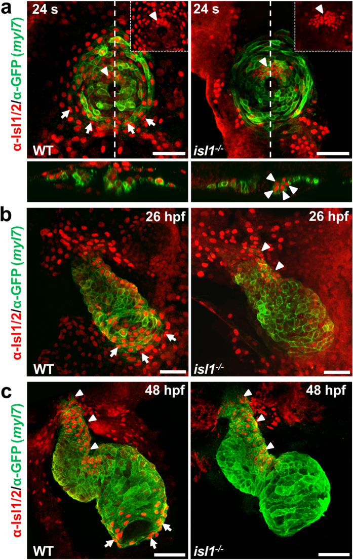
Figure 1. Residual Isl1/2 positive cells in isl1−/− zebrafish hearts.
(a–c) Confocal images of wild-type sibling and Tg(myl7:EGFP-HsHRAS)s883 isl1−/− embryos stained with anti-GFP and anti-Isl1/2 antibodies at 24 somites (a), 26 hpf (b) and 48 hpf (c). Arrows point to Isl1+ cells at the periphery of the cone (a) or Isl1+ cardiomyocytes at the venous pole of the atrium (b,c), arrowheads point to residual Isl1/2+ cardiomyocytes in the future ventricle (a) or the inner curvature of the ventricle and the outflow pole (b,c). Scale bars, 50 μm.

