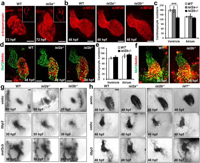
Figure 3. Isl2b-deficiency leads to defects in anterior SHF development.
(a) Confocal images of control and isl2a−/− hearts stained with anti-tropomyosin antibody at 72 hpf, showing impaired displacement of the ventricle towards the right side. (b) Confocal images of control, isl2a−/− and isl2b−/− embryos stained with anti-MF20 antibody at 48 hpf. Isl2a−/− hearts were imaged from the side to analyze the role of isl2a in heart chamber formation. Scale bars in (a,b), 50 μm. (c,d) Number of atrial and ventricular cardiomyocytes (c) quantified following whole mount immunostaining with anti-Mef2 and anti-Amhc antibody (S46) of wild-type, isl2a−/− and isl2b−/− embryos at 48 hpf (d). (e,f) Number of atrial and ventricular cardiomyocytes (e) quantified following whole mount immunostaining with anti-Mef2 and anti-Amhc antibody (S46) of wild-type and isl2b−/− embryos at 30 hpf (f). (g) In situ hybridization for vmhc, ltbp3 and mef2cb of control, isl2a−/− and isl2b−/− embryos at 30 hpf. (h) In situ hybridization for vmhc, amhc and ltbp3 of control, isl2a−/−, isl2b−/− and isl1−/− embryos at 48 hpf. Data information: In (c), data are presented as mean ± SEM. ***p < 0.001 (Student’s t-test).

