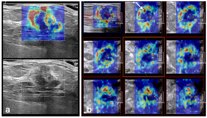
Figure 1. Invasive ductal carcinoma grade 2 in a 61-year-old woman.
The 2D B-mode image shows an irregular hypoechoic mass considered to be BI-RADS category 4b (bottom). On 2D SWE (a) and 3D SWE (b), an ROI for measuring the ESDE value was placed to include the whole breast mass and immediately adjacent stiff tissue. The other Q-Box for measuring the ESDU was placed within the mass to encompass the maximum portion of the mass and exclude tissue outside the mass. (a) Emax value recorded using the larger Q-Box. (b) Emax value recorded using a 2-mm2 Q-Box (arrow).

