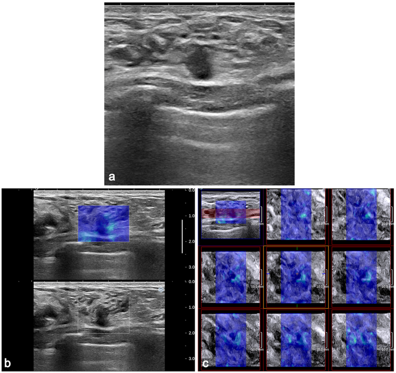
Figure 4. DCIS in a 36-year-old woman.
(a) The 2D B-mode image shows a 7.4-mm, irregular hypoechoic mass with an uncircumscribed margin considered to be BI-RADS category 4a. Both 2D SWE (b) and 3D SWE (c) show a homogeneously soft (blue colour) mass and surrounding tissue (2D Emax 58.7 kPa, ESDE 10.2 kPa, ESDU 9.4 kPa; 3D Emax 87.1 kPa, ESDE 11.8 kPa, ESDU 9.5 kPa). When combining SWE with B-mode US, BI-RADS category 3 was assigned to the mass.

