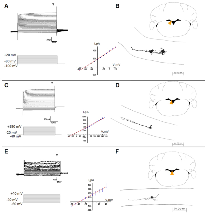
Figure 6. Electrophysiological characteristics of cells of the SVCC.
Whole-cell patch-clamp sample recordings from cells at different locations in the SVCC (orange arrowheads in coronal cerebellar slices). Different current profiles were dissected after application of the protocols indicated (A–C and E). Recordings in (A–C) correspond to glial cells, while E is similar to those of neuroblasts of the lateral ventricles. Notice the linear current-voltage relation (I–V plots); these were calculated at the end of the current steps (gray arrowheads). During the recordings, the cells were filled with biocytin. The current profiles correlate with diverse cell morphologies unveiled by drawing on paper the biocytin labeled cells, using a camera lucida (B, D–F). Cells in B were located in the lateral portion of the SVCC and possess large soma (20–25 μm), short projections and one long processes that projects laterally. In D, cells with smaller soma (5–8 μm) and two long lateral processes; in F, cells were distributed medially, with small soma (8-10 μm) and multiple projections extended transversally in the medial portion of the SVCC.

