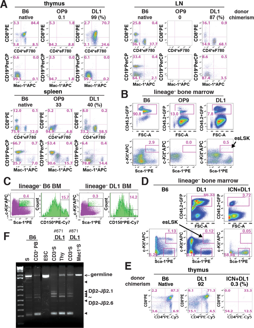
Figure 2. Notch-activated ESC-HSC-DL1 Specifies to esLSKs Upon Engraftment.
(A) Donor-derived reconstitution of standard (OP9) and Notch-activated (DL1) AV15 ESC-HSCs in the thymus, lymph nodes (LN) and spleen of representative NSGs at 12 wks post-transplant. In each panel, percent donor chimerism (CD45.2+ and GFP+), donor-derived CD4/CD8 (middle) and CD19/Mac-1 (bottom) lineages are shown. (B–C) LSK-CD150 profile of BM-engrafted AV15 ESC-HSCs. esLSKs are indicated by arrow. (D–E) BM and thymic engraftment of DL1- and ICN+DL1-activated iICN ESC-HSCs at 16 wks post-transplant. Arrow indicates esLSKs. (F) PCR analysis of TCRβ-DJ rearrangement in sorted lineages of two representative iICN ESC-HSC-DL1 recipients at 16 wks post-transplant. Arrow: germline amplicons; triangles: Dβ2-Jβ2.1 to Dβ2-Jβ2.6 amplicons. Lane#1-2: B6 genomic DNA of splenocytes (S) and peripheral CD3+ T cells (CD3+ PB); #3: ESC; #4-7: CD3/Mac-1+ splenocytes and total thymocytes. See also Fig.S3.

