Fig. 2.

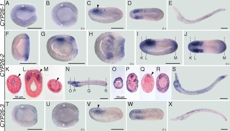
Developmental expression patterns of amphioxus CYP26 genes. Whole mount in situ hybridization experiments were carried out for CYP26-1 (a-e), CYP26-2 (f-s), and CYP26-3 (t-x). Amphioxus (Branchiostoma lanceolatum) embryos and larvae are shown as lateral views with anterior to the left and the dorsal side up, excepting for (b, d, h, j, u, w), which are dorsal views (d.v.) with anterior to the left, and for (k-m) and (o-r), which are cross-sections viewed from the front. Cross-sections in (k-m) are through the embryo shown in (i, j) at the levels indicated by the dashed lines and cross-sections in (o-r) are through the embryo shown in (n) at the levels indicated by the dashed lines. White asterisks in (a, b, t, u) highlight inconspicuous early expression domains of CYP26-1 and CYP26-3. Arrowheads in (c, v) indicate central nervous system expression and in (k-m, q) ectodermal signal. Developmental stages shown are: early gastrula (f), late gastrula (a, b, g, h, t, u), mid neurula (c, d, i-m, v, w), very early larva (n-r), early (60 h) larva (e, s, x). Scale bars are 100 μm for the whole mounts and 50 μm for the cross-sections
