Figure 6.
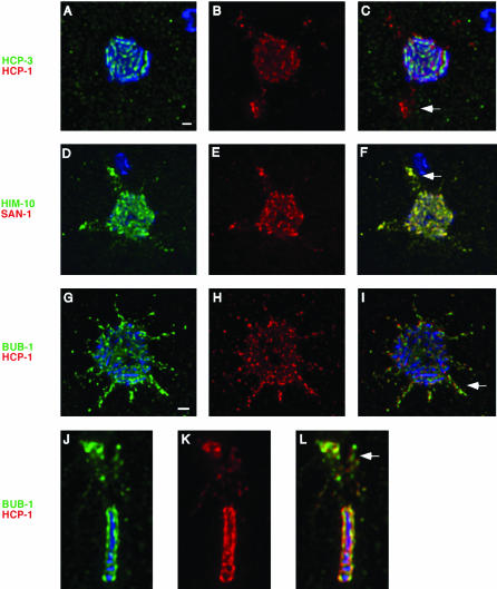
The C. elegans kinetochore exhibits a distinct reorganization in response to checkpoint stimuli. After exposure to either anoxia (a-f, j-l) or nocodazole (g-i), embryos were stained with antibodies against HCP-3/CeCENP-A (a-c), HIM-10/CeNUF2 (d-f), and BUB-1 (g-l) (green), as well as HCP-1/CeCENP-F (a-c, g-l) and SAN-1/CeMAD3 (d-f) (red). Metaphase plates in four cell embryos were examined to determine the effect that checkpoint activation would have on kinetochore organization. The centromere did not change in response to this treatment as seen by HCP-3/CeCENP-A staining (a). However, the kinetochore underwent significant rearrangements after exposure to either nocodazole or anoxia. This was characterized by the localization of all of the kinetochore proteins in large flares projecting out from the metaphase plate (b-i). The flares also could be detected when metaphase plates were observed from the perpendicular perspective (j-l), appearing as aggregates of staining lying above or below the metaphase plate. White arrows in the merged panels highlight the location of the flares. Bar, 1 μm.

