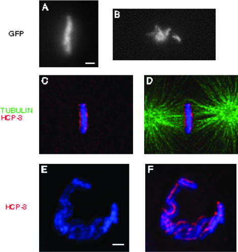
Figure 7.
hcp1/hcp2(RNAi) and him-10(RNAi) embryos exhibit defects at metaphase. In the histone H2B::GFP background, wild-type (a) and hcp1/hcp2(RNAi) (b) chromosomes were visualized in one cell embryos shortly after pronuclear fusion. We filmed the progression of these embryos from prometaphase to anaphase (see Supplemental Figure 2), and the frames shown here represent the maximum level of congression observed in these backgrounds. Whereas wild-type chromosomes congress to the center of the cell and align properly at metaphase (a), hcp1/hcp2(RNAi) chromosomes fail to form a metaphase plate. him-10(RNAi) (c-f) embryos were stained with DAPI (blue), α-tubulin (green), and α-HCP-3 (red). Whereas him-10(RNAi) chromosomes are able to congress to the center of the cell, as seen from the perpendicular perspective (c and d), they do not organize into a proper metaphase plate, as seen from the centrosomal perspective (e and f). Instead of the solid plate observed in wild-type (see Figure 2), the chromosomes assemble into an open, horseshoe-shaped structure that is indicative of a failure to organize metaphase chromosomes. Bar, 1 μm.

