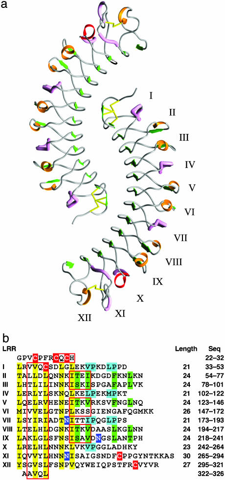
Fig. 1.
Structure of the LRR domain of bovine decorin. (a) Ribbon diagram of the dimeric structure of decorin LRR domain. Green arrows, β-strands; red ribbons, α-helical turns; pink tubes, segments of polyproline II helix; orange ribbons, short segments of 310 helices and β-turns; yellow sticks, disulfide bonds. (b) Internal organization of bovine decorin LRRs (residues 22-326). Yellow highlight, LRR consensus residues; red highlight, Cys residues; green highlight, consensus residues for the 24-aa repeat; cyan highlight, consensus residues for the 21-aa repeat; blue highlight, Asn residues with oligosaccharide substituents; red boxes, amino acids that contribute to β-sheets.

