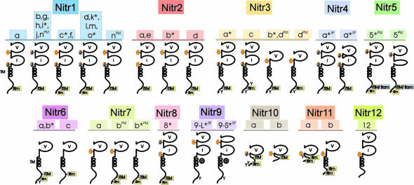
Fig. 2.
Schematic representation of diverse NITRs in zebrafish. Predicted protein structures of multiple zebrafish NITRs are shown. Protein domains include the following: a variable Ig domain (V); an intermediate Ig domain (I) (11); a transmembrane region (TM) denoted by a helical structure; a positively charged residue midmembrane ( ); a prototypic joining-like region including the GXGTXLX(V/I/L) peptide sequences (J); a J-like variant (see Table 3) is indicated by an orange oval; a conventional ITIM including the (I/V/L)XYXX(I/V/L) peptide sequences; a variant of the conventional ITIM (itim); and variant of the conventional ITAM sequence (itam). An asterisk (*) indicates that full-length cDNA clones have been identified and sequenced confirming these structures; other structures are predicted from genomic sequence. SP, alternative RNA splicing variants; PM, polymorphic variants of the same gene.

