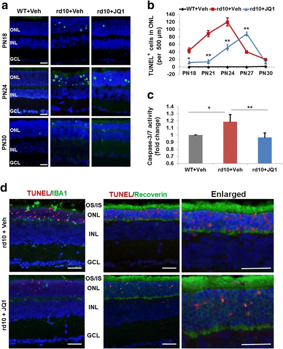
Fig. 2.

JQ1 treatment inhibits apoptosis in the rd10 mouse retina. Intravitreal injection of JQ1 (or vehicle) was performed at PN14, as described in Fig. 1. Eyeballs were collected at PN18, PN21, PN24, PN27, and PN30. Cryosections were prepared and used for TUNEL staining. a Representative TUNEL (green) images showing an inhibitory effect of JQ1 in rd10 retinas. Scale bar 50 μm. Blue: DAPI staining of nuclei. For images of PN21 and PN27, see Additional file 1: Figure S2. b Quantification of TUNEL-positive cells (per 500 μm ONL length): mean ± SEM, n = 6 mice; **P < 0.01, *P < 0.05 compared to rd10 vehicle control. c Caspase-3/7 activity assay showing an inhibitory effect of JQ1 on retinal cell apoptosis. For the assay, homogenates were prepared from retinas collected and pooled from six mice at PN24. **P < 0.01 compared to rd10 vehicle control, n = 3 independent assay experiments. d For TUNEL/IBA1 and TUNEL/Recoverin co-staining (PN24 retinas), a TMR red kit (Roche) was used so that TUNEL-positive nuclei appear red. The data show that TUNEL-positive nuclei do not overlap with IBA1-positive cells; instead, they are localized within recoverin-positive (photoreceptor) cells. Scale bar 50 μm
