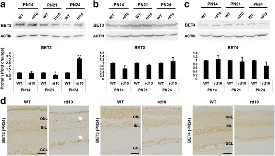
Fig. 6.

BET2 is upregulated in the rd10 mouse retina compared to B6 WT control. a–c Western blots of retinal homogenates collected from B6 or rd10 mice at PN14, P21, or PN24 (>6 mice at each time point). Quantification: normalization to β-actin and WT at each time point; mean ± SEM; n = 3 independent Western blot experiments; *P < 0.05, **P < 0.01 compared to WT control. d Immunostaining of BET2, BET3, and BET4 on paraffin-embedded retinal sections collected at PN24. Arrow points to dots of condensed staining. Scale bar 50 μm
